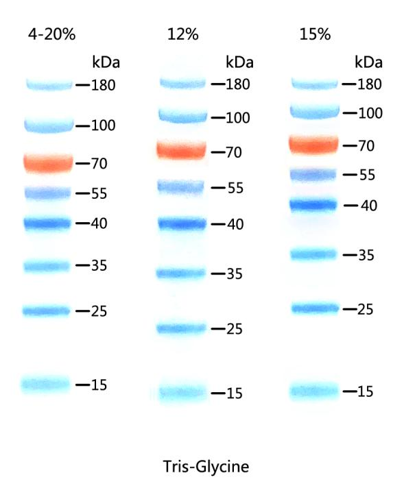
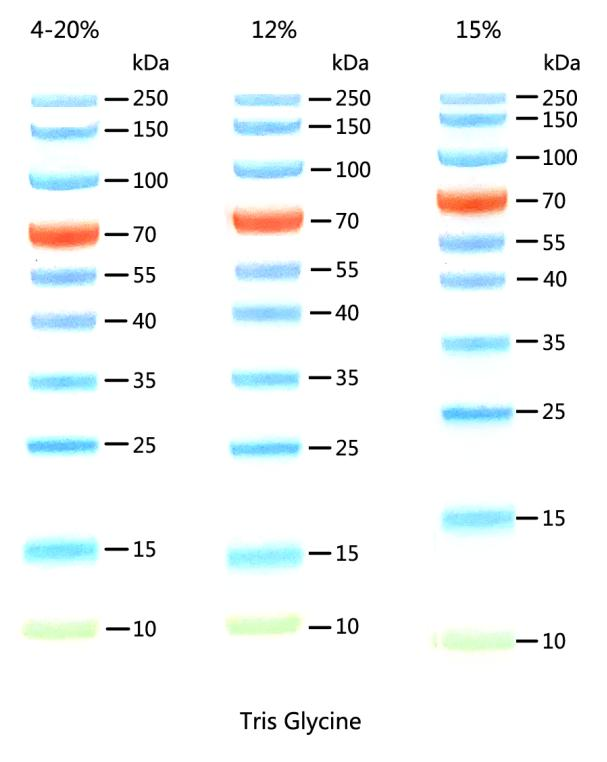
10-180KD Prestained Protein Ladder D1013-A D1013-B
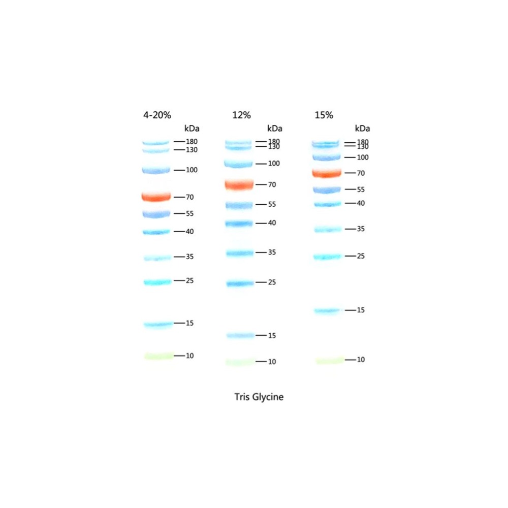
10-180KD Prestained Protein Ladder D1013-A D1013-B
10-180KD Prestained Protein Ladder
Cat. No./Spec.
D1013-A/250 μl; D1013-B/250 μl×5
Component
Components | D1013-A | D1013-B |
10-180KD Prestained Protein Ladder | 250 μl | 250 μl×5 |
Storage
-20 ̊C.
Description
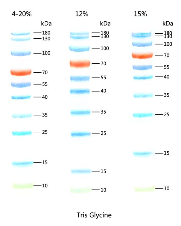
This product contains 10 pre-stained standard proteins with known molecular weights, ranging from 10kDa to 180kDa. The 70kDa band is marked with an orange protein band, and the 10kDa band is marked with a green protein band, while the others are blue. It allows for direct observation of the protein electrophoresis process and a clear assessment of the Western blot transfer effect. Typically, a sample volume of 5µl per application is adequate.
Bands (kDa)
10, 15, 25, 35, 40, 55, 70, 100, 130, 180
Migration Patterns
Application:
For SDS-PAGE, Western Blot.
Monitor the electrophoresis process throughout,
Assess the efficiency of transfer,
Precisely locate the target protein.
Features:
Ready-to-use, multicolor pre-stained
Bright colors, clear bands
High purity protein, accurate molecular weight
Multiple bands, wide range
Good consistency between batches
Prestained Protein Ladder
| 15-180KD Prestained Protein Ladder
| 10-250KD Prestained Protein Ladder | 10-180KD Prestained Protein Ladder
| 10-250KD Prestained Immunoblotting Protein Ladder
|
Molecular weight range | 15-180 kDa | 10-250 kDa | 10-180 kDa | 10-250 kDa |
Band quantity | 8 | 10 | 10 | 12 |
Band molecular weight (kDa) | 15, 25, 35, 40, 55, 70, 100, 180 | 10, 15, 25, 35, 40, 55, 70, 100, 150, 250 | 10, 15, 25, 35, 40, 55, 70, 100, 130, 180 | 10, 15, 25, 30, 35, 40, 55, 70, 80, 100, 150, 250 |
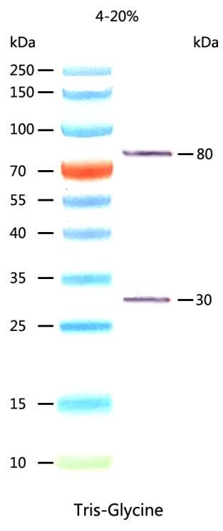
Color | Blue, orange | Blue, orange, green | Blue, orange, green | Blue, orange, green. IgG binding sites are located on 2 bands (80 and 30 kDa). |
Imaging method | Visual color comparison
| Visual color comparison | Visual color comparison | Visual color comparison. bands at 80 and 30 kDa can be visualized by Western Blot and Coomassie Brilliant Blue staining. |
Recommended gel system | Tris-Glycine | Tris-Glycine,MOPS | Tris-Glycine | Tris-Glycine |
Cat. No. / Spec. | D1011-A/250 μl D1011-B/250 μl×5 | D1012-A/250 μl D1012-B/250 μl×5 | D1013-A/250 μl D1013-B/250 μl×5 | D1014-A/250 μl D1014-B/250 μl×5 |
 Tel: +86 20 31600213
  Sales EMail: order@gdsbio.com

