Tel: +86 20 31600213 Tel: +86 20 31600213  Sales EMail: order@gdsbio.com Sales EMail: order@gdsbio.com |
Search
|

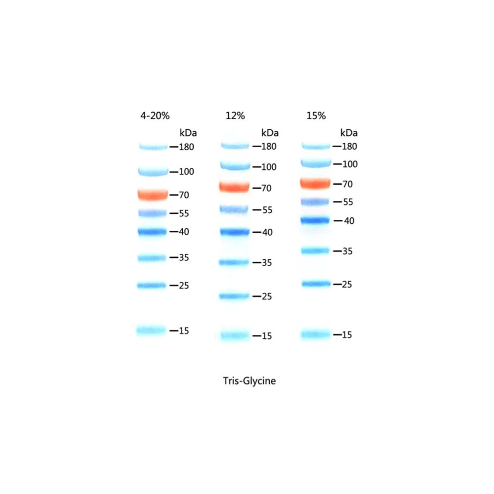
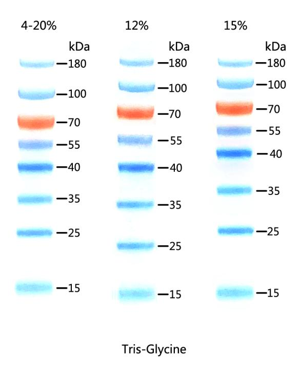
15-180KD Prestained Protein Ladder D1011-A D1011-B
15-180KD Prestained Protein Ladder D1011-A D1011-B
15-180KD Prestained Protein Ladder
Cat. No./Spec.
D1011-A/250 μl; D1011-B/250 μl×5
Component
Components | D1011-A | D1011-B |
15-180KD Prestained Protein Ladder | 250 μl | 250 μl×5 |
Storage
-20 ̊C.
Description
This product contains 8 pre-stained standard proteins with known molecular weights, ranging from 15kDa to 180kDa. The 70kDa band is marked with an orange protein band, while the others are blue. It allows for direct observation of the protein electrophoresis process and a clear assessment of the Western blot transfer effect. Typically, a sample volume of 5µl per application is adequate.
Bands (kDa)
15, 25, 35, 40, 55, 70, 100, 180
Migration Patterns
Application:
For SDS-PAGE, Western Blot.
Monitor the electrophoresis process throughout,
Assess the efficiency of transfer,
Precisely locate the target protein.
Features:
Ready-to-use, multicolor pre-stained
Bright colors, clear bands
High purity protein, accurate molecular weight
Multiple bands, wide range
Good consistency between batches
Prestained Protein Ladder
| 15-180KD Prestained Protein Ladder
| 10-250KD Prestained Protein Ladder | 10-180KD Prestained Protein Ladder
| 10-250KD Prestained Immunoblotting Protein Ladder
|
Molecular weight range | 15-180 kDa | 10-250 kDa | 10-180 kDa | 10-250 kDa |
Band quantity | 8 | 10 | 10 | 12 |
Band molecular weight (kDa) | 15, 25, 35, 40, 55, 70, 100, 180 | 10, 15, 25, 35, 40, 55, 70, 100, 150, 250 | 10, 15, 25, 35, 40, 55, 70, 100, 130, 180 | 10, 15, 25, 30, 35, 40, 55, 70, 80, 100, 150, 250 |
Color | Blue, orange | Blue, orange, green | Blue, orange, green | Blue, orange, green. IgG binding sites are located on 2 bands (80 and 30 kDa). |
Imaging method | Visual color comparison
| Visual color comparison | Visual color comparison | Visual color comparison. bands at 80 and 30 kDa can be visualized by Western Blot and Coomassie Brilliant Blue staining. |
Recommended gel system | Tris-Glycine | Tris-Glycine,MOPS | Tris-Glycine | Tris-Glycine |
Cat. No. / Spec. | D1011-A/250 μl D1011-B/250 μl×5 | D1012-A/250 μl D1012-B/250 μl×5 | D1013-A/250 μl D1013-B/250 μl×5 | D1014-A/250 μl D1014-B/250 μl×5 |


Guangzhou Dongsheng Biotech Co., Ltd. Web: www.gdsbious.com Email: order@gdsbio.com WhatsApp/Wechat: +86 19128993865 TEL: 86-020-31600213 ADD: Room 305, Building A, No. 179, Guangpu East Road, Guangzhou 510760, China | Recommend |